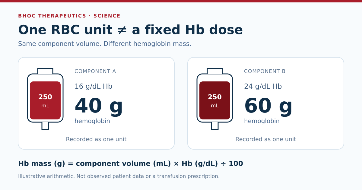
A transfusion record may show two units of red blood cells. That count does not establish the exact mass of hemoglobin administered. It also does not establish how much oxygen reached the patient’s tissues.
Before comparing transfusion results, we should ask what was standardized, what was actually infused, when the response was measured and which function improved. These questions apply to donor blood, red blood cell components and the development of oxygen therapeutics.
The hemoglobin dose inside the bag
Agnihotri and colleagues measured hemoglobin content in 125 RBC units and found a range of 42.3 to 80.8 g per unit. Jain and colleagues examined 900 packed RBC units and reported 30.77 to 87.36 g, with a mean of 52.91 ± 9.99 g. The latter study also reported component volumes of 155 to 370 mL. [1,2]
| Study | Units tested | Hb per unit | Maximum / minimum |
|---|---|---|---|
| Agnihotri 2014 | 125 | 42.3 to 80.8 g | 1.91 |
| Jain 2022 | 900 | 30.77 to 87.36 g | 2.84 |
These are ranges from specific studies, not universal limits for every blood service. Different collection volumes and processing conditions contributed to the variation. Even so, they demonstrate why the number of bags alone is an incomplete description of hemoglobin dose. [1,2]
For illustration, a 250 mL component containing 16 g/dL Hb holds 40 g of Hb. The same volume at 24 g/dL holds 60 g. Both can be recorded as one unit, despite a 50% difference in Hb mass. This is an arithmetic example, not a proposed transfusion prescription.
What blood component standards specify
Blood components have defined quality specifications. For example, the UK JPAC specification for Red Cells in Additive Solution, Leucocyte Depleted includes the parameters below. [3]
| Parameter | UK JPAC specification |
|---|---|
| Component volume | 280 ±60 mL |
| Hemoglobin content | At least 40 g per unit |
| Hematocrit | 0.50 to 0.70 L/L |
| Hemolysis | Less than 0.8% of red cell mass |
These are specifications with sampling, conformity and concessionary-release provisions, not identical values for every bag. Hb content is monitored in 1% of components or through statistical process control, with every available component tested when production is no more than 10 per month. [3]
JPAC also permits a calculated or nominal labelled volume. For RBC components, that volume includes the anticoagulant and any additive solution. It therefore describes the whole component, not its red cell volume or Hb mass alone. [4]
Compliance with a component specification is valuable information. It does not, by itself, provide a measured Hb dose and oxygen-delivery potency for each unit administered.
Agostini and colleagues’ 2025 European validation of RBCs processed with Hemanext ONE provides a practical example. The study assessed Hb content, hematocrit and hemolysis against acceptance criteria during storage. Table 3 reported mean Hb content after processing of 50.3 ± 5.0 g/unit in Bergen and 55.2 ± 5.0 g/unit in Genoa. Successful process validation therefore coexisted with variation in measured unit contents. This was an in vitro validation, not a comparison of clinical outcomes. [16]
Donor biology and processing change the component
In an international survey, Shih and colleagues collected data from 10 blood centres in nine countries. Collection and processing differed in cooling, holding time, donor Hb limits, separation methods, leucoreduction timing, equipment and shelf life. Quality testing also varied. The study identified differences in laboratory characteristics, without establishing that one production method produced better clinical outcomes. [5]
A component name can therefore cover products made under different conditions. Comparisons should identify the component and its processing history, rather than treating every RBC unit as the same preparation.
Oxygen exposure during storage is another variable. Yoshida and colleagues analyzed oxygen saturation within 24 hours of collection in 492 leucocyte-reduced RBC units in AS-3. Mean saturation was 45.9 ± 17.5%, demonstrating substantial initial variation. A separate pool-and-split experiment linked higher storage oxygen saturation with less favorable metabolic and oxidative quality markers. These were measurements in blood components, not measurements of patient oxygenation. The study identifies an additional source of product variability; it does not establish a clinical benefit from controlling it. [17]
Regulatory status does not mean identical composition
The regulatory position depends on the jurisdiction. In the United States, FDA regulates blood components as drugs and biological products. Blood establishments generally require registration and product listing, subject to exemptions. Licensed blood products exist, and FDA describes biologics licence applications as a route for approval of blood components. A blanket description of blood or RBCs as unregistered products would be incorrect. [6,7]
In the European Union, Article 3(6) of Directive 2001/83/EC excludes whole blood, plasma and blood cells of human origin from that medicinal-products directive, with an exception for industrially prepared plasma. Transfusion components are covered by dedicated blood quality and safety requirements, including Directive 2002/98/EC. The SoHO Regulation (EU) 2024/1938 generally applies from 7 August 2027. National rules also matter. [8-10]
Registration, product licensure, establishment authorization and component release describe different regulatory steps. None should be used as a substitute for describing the measured composition and function of the administered component.
Patient hemoglobin is one part of oxygen delivery
The Hb concentration reported by a patient’s blood test is different from the Hb mass in the transfused component. Concentration depends on the amount of hemoglobin relative to circulating blood volume. During resuscitation, ongoing bleeding and fluid administration can change that relationship. [11]
A rise in Hb documents a change in concentration. Tissue oxygenation also depends on blood flow, oxygen saturation, oxygen release and distribution through the microcirculation. Our scientific approach therefore distinguishes oxygen-carrying capacity from delivery and the response of tissues. [12]
These distinctions help explain why a post-transfusion Hb value should be interpreted alongside the patient’s physiological state. They also prevent a laboratory concentration from being presented as direct proof that organ oxygenation has recovered.
What a before and after comparison can tell us
Our methodological proposal is to report the intervention as fully as possible: component identity, volume actually infused, Hb content per unit where measured, total administered Hb mass, storage age and relevant processing. Any calculated or assumed value should be labelled as such.
The observation period matters. A sample collected during active bleeding answers a different question from one collected after source control and fluid redistribution. Concurrent crystalloids, plasma, platelets and vasoactive treatment should be recorded when interpreting the response.
Reuland and colleagues’ 2026 ICU analysis illustrates the problem. They found considerable variation across four public datasets and explicitly identified variation in RBC unit volume and Hb content as a limitation of blood-loss estimation. Source-control information was also insufficient. Their findings describe practice variation; they do not identify the best treatment strategy. [11]
For research, the measurement should match the intended claim. The table below separates the administered dose from laboratory function and the patient’s response.
| Measurement | What it addresses |
|---|---|
| Administered volume and Hb mass | Amount of component and Hb delivered to the patient |
| Oxygen loading and unloading assays | Function under specified laboratory conditions |
| Hb, perfusion and oxygenation measurements | Patient response at defined times and sites |
| Organ function, adverse events and survival | Clinical benefit and harm in the studied setting |
Storage and functional potency need separate assessment
Rogers and colleagues proposed lung-to-tissue oxygen flux, L-TOF, as an in vitro metric combining oxygen association and dissociation behaviour. In their model, 476 ± 21.6 mL of day-42 RBC concentrate stored in AS-1 was calculated to match 300 mL of fresh RBC concentrate. That is approximately 1.59 times the volume. [13]
This is a modeled potency comparison. It is not a bedside dosing instruction or proof of a clinical outcome difference. The method does not replace assessment of vascular response, microcirculation, safety or patient outcomes. [13]
Clinical evidence must be considered separately. In the ABLE randomized trial, 2,430 critically ill adults received either fresh RBCs or standard-issue RBCs. Fresh blood did not reduce 90-day mortality. This finding limits any broad claim that shorter storage alone improves survival in that population. [14]
What oxygen-controlled storage studies actually measure
The original studies cited by Hemanext help distinguish several levels of evidence. In 2022, Rabcuka and colleagues used microfluidic imaging to measure oxygen unloading from individual RBCs. Hypoxic storage preserved faster unloading in samples stored for up to 35 days. This demonstrates a laboratory functional difference under the tested conditions. It does not show how much additional oxygen reaches a patient’s organs. [18]
D’Alessandro and colleagues studied donor-paired units from 12 healthy volunteers, stored conventionally or under hypoxia for 42 days. Hypoxically stored RBCs showed better autologous post-transfusion recovery, with comparable hemolysis. This measures how many transfused cells remain in circulation. Cell recovery is relevant to the effective circulating dose, but it is a different endpoint from tissue oxygenation or clinical benefit in bleeding patients. [19]
Rabcuka and colleagues subsequently tested stored RBCs in ex vivo perfused human kidneys. Switching from conventionally to hypoxically stored RBCs reversibly increased renal respiratory rate by approximately 20% and estimated oxygen diffusion capacity by approximately 70%. These findings connect RBC storage conditions with oxygen transfer in living human tissue outside the body. They do not establish improved transplant outcomes or the same effect after transfusion into a patient. The paper appeared online in 2025 and in the 2026 journal volume. [20]
A 2026 clinical pilot by Reikvam and colleagues brings the measurement question directly to patients. Ten patients with hematological malignancies received two hypoxic RBC units each; ten patients undergoing burn excision received a mean of 1.8 ± 0.4 units. Hb was checked 15 to 60 minutes after transfusion. Mean increases were 1.2 ± 0.5 g/dL and 0.3 ± 1.0 g/dL, respectively. These groups had different clinical circumstances and received different amounts. The findings illustrate why an Hb increment must be interpreted with dose, timing and clinical context. [21]
No adverse events were attributed to the hypoxic RBC transfusions in that pilot. The study was small, open-label and non-comparative. It provides preliminary safety observations, not proof of superior efficacy, reduced transfusion requirements or improved tissue oxygenation. [21]
Our interpretation is that administered Hb mass, circulating cell recovery and oxygen-transfer function should be characterized separately. Hemanext processes donor RBCs; these findings concern the tested RBC storage systems. They cannot be transferred directly to a cell-free hemoglobin formulation or to BHOC.
Uncertain dose narrows the conclusions we can draw
When Hb content is not measured per unit, the result remains observable. The patient’s Hb, physiological measurements and clinical outcomes can still be recorded reliably. The uncertainty concerns how that response relates to the exact administered dose.
Our interpretation is that a well-designed randomized trial can estimate the effect of a transfusion policy even when individual components vary. A study comparing unit-based policies answers a policy question. It does not automatically establish a precise relationship between grams of Hb and tissue oxygenation.
Atilla and colleagues tested Hb-content-based selection in 89 patients across 178 randomized transfusion episodes. The authors reported 19.8% fewer units than originally ordered in the study arm, with no statistically significant difference in achieving target Hb. This supports further work on dose characterization; it does not establish equivalent survival or tissue-oxygenation outcomes. [15]
Knowing the administered Hb mass would reduce one source of uncertainty. Recipient physiology and the clinical setting would still need to be considered.
The same requirement applies to BHOC
For BHOC, Biological Hemoglobin Oxygen Carrier, these observations define a development requirement. A formulation intended for Precision Oxygen Therapeutics should have a characterized dose, reproducible manufacturing specifications and measured functional properties. [12]
We believe that analytical oxygen handling should be linked to direct assessment of vascular behaviour, tissue oxygenation and clinical benefit in the intended use. Predictability must be demonstrated for the specific formulation and its storage conditions. Findings about donor RBC variability do not establish the efficacy or safety of BHOC.
Comparisons should state the exact products, administered doses, measurement times and endpoints. That is how an observed response can become an interpretable treatment effect.
Sources
- Agnihotri N, Pal L, Thakur M, Kumar P. The need to label red blood cell units with their haemoglobin content: a single centre study on haemoglobin variations due to donor-related factors. Blood Transfus. 2014;12:520-526. Original source ↗
- Jain R, Sachdev S, Marwaha N, Gupta A. Assessment of Hemoglobin Content of Packed Red Cells: A prospective Study on Hemoglobin Content Variation Due to Donor, Collection, and Processing-Related Factors. Is it Time to Label Each Unit with Hemoglobin Content? Glob J Transfus Med. 2022;7:36-41. Original source ↗
- JPAC. Red Book, section 7.3.2. Red Cells in Additive Solution, Leucocyte Depleted. Specification and testing provisions. Original source ↗
- JPAC. Red Book, section 6.3. Component and process monitoring tests. Sampling, monitoring and labelled volume. Original source ↗
- Shih AW, Apelseth TO, Cardigan R, et al. Not all red cell concentrate units are equivalent: international survey of processing and in vitro quality data. Vox Sang. 2019;114:783-794. Original source ↗
- US FDA. FDA Regulation of Blood and Blood Components in the United States. Regulatory education material. Original source ↗
- US FDA. Blood Establishment Registration and Product Listing. Original source ↗
- European Parliament and Council. Directive 2001/83/EC, Article 3(6). Community code relating to medicinal products for human use. Original source ↗
- European Parliament and Council. Directive 2002/98/EC. Quality and safety requirements for human blood and blood components. Original source ↗
- European Parliament and Council. Regulation (EU) 2024/1938. Quality and safety standards for substances of human origin intended for human application. Article 87. Original source ↗
- Reuland MC, Raasveld SJ, Dongelmans DA, Elbers PWG, Müller MCA, Vlaar APJ. Hemorrhagic shock in intensive care: Variation in blood product administration during resuscitation. Vox Sang. First published 23 September 2026. Original source ↗
- BHOC Therapeutics. Scientific Evidence Relevant to BHOC and Oxygen Therapeutics. Physiological context and development positioning. Original source ↗
- Rogers SC, Brummet M, Tobin KV, et al. Defining and quantifying oxygen delivery potency of blood products. Blood Red Cells & Iron. 2026;2(2):100054. Original source ↗
- Lacroix J, Hébert PC, Fergusson DA, et al. Age of transfused blood in critically ill adults. N Engl J Med. 2015;372:1410-1418. Original source ↗
- Atilla E, Toprak SK, Civriz Bozdağ S, Topçuoğlu P, Arslan Ö. A Randomized Comparison of Hemoglobin Content-Based Versus Standard (Unit-Based) Red Blood Cell Transfusion Policy. Turk J Haematol. 2017;34:244-249. Original source ↗
- Agostini V, Henschler R, Felli Lunde TH, et al. A validation study of the in vitro performance of hypoxic red blood cells for transfusion across centers in Europe. Transfusion. 2025;65:2306-2315. Quantitative comparisons here use the timed measurements in Table 3. Original source ↗
- Yoshida T, Blair A, D’Alessandro A, et al. Enhancing uniformity and overall quality of red cell concentrate with anaerobic storage. Blood Transfus. 2017;15:172-181. Original source ↗
- Rabcuka J, Blonski S, Meli A, et al. Metabolic reprogramming under hypoxic storage preserves faster oxygen unloading from stored red blood cells. Blood Adv. 2022;6:5415-5428. Original source ↗
- D’Alessandro A, Yoshida T, Nestheide S, et al. Hypoxic storage of red blood cells improves metabolism and post-transfusion recovery. Transfusion. 2020;60:786-798. Original source ↗
- Rabcuka J, Fallon J, Meli A, et al. Storage under hypoxia improves the ability of red cells to release oxygen in ex vivo perfused human kidneys. Blood Red Cells & Iron. 2026;2(1):100038. Published online in 2025. Original source ↗
- Reikvam H, Felli Lunde TH, Kristoffersen EK, et al. A Clinical Investigation of Hypoxic Red Blood Cell Administration in Patients with Transfusion-Dependent Hematological Malignancies and Burns. J Blood Med. 2026;17:573232. Original source ↗